
Persistent Concussion Symptoms: Are We Assessing Everything the Neck Actually Does?
An athlete is four weeks post-concussion.
Their recovery has plateaued. They continue to experience headaches, dizziness, imbalance, visual complaints, or difficulty progressing back to sport.
At this point, it is easy to assume the remaining symptoms are still coming from the brain.
But here's a question worth asking:
What are the chances the neck was injured too?
The forces involved in concussion do not necessarily affect the brain in isolation. Rapid acceleration, deceleration, and rotation of the head also place considerable demands on the cervical spine.
If enough force was transmitted to produce a concussion, is it reasonable to assume the neck escaped injury?
The challenge is that cervical dysfunction doesn't always present as neck pain alone. Impaired cervical sensorimotor function may affect the quality of information reaching the brain about head position and movement, potentially contributing to dizziness, imbalance, visual complaints, and difficulty coordinating movement.
Before assuming ongoing symptoms are simply "the concussion," perhaps we need to look a little lower. Cheever (2016) stated: If patients exhibit dizziness, headache, or other symptoms after a collision, they are almost automatically diagnosed as having sustained a concussion. However, patients with cervical injury after a pathomechanical event affecting the head or neck may manifest nearly identical symptoms.
Are We Assessing the Neck Like Every Other Joint?
Most physiotherapists are very comfortable assessing the cervical spine.
We assess range of motion and use palpation to evaluate things such as muscle tenderness, joint tenderness, and joint mobility.
These findings are important.
But imagine assessing a lateral ankle sprain the same way.
Would you assess ankle range of motion and palpatory findings, and then stop?
Of course not.
We routinely assess strength, proprioception, balance, motor control, and functional performance because we recognize that restoring pain-free movement alone doesn't mean the ankle has fully recovered.
Do we routinely apply the same standard to the cervical spine?
The cervical spine isn't simply a collection of joints and muscles. It is also an extraordinarily rich source of proprioceptive information.
The small suboccipital muscles have a particularly high concentration of muscle spindles. One anatomical study reported approximately 242 muscle spindles per gram in the inferior oblique suboccipital muscle compared with only 2.2 per gram in the trapezius. Although direct comparisons of spindle density between muscles of very different sizes should be interpreted cautiously, these findings illustrate just how richly innervated the upper cervical region is for proprioception.
That makes sense. The nervous system needs extremely precise information about the position of the head relative to the trunk. Cervical proprioceptive information integrates with visual and vestibular input to contribute to gaze stability, balance, postural control, and coordinated movement.
If that system has been affected by injury, range of motion and palpation alone may not tell us the whole story.
Could Cervical Sensorimotor Dysfunction Matter?
Research has identified abnormalities in cervical joint position sense, head repositioning accuracy, cervical movement control, and muscle activation in people with cervical disorders, with emerging evidence also identifying cervical sensorimotor changes following concussion.
This doesn't mean cervical dysfunction explains every persistent symptom. It is simply one potentially modifiable contributor that shouldn't be overlooked.
Consider what happens when an athlete returns to the field, court, or ice.
They need to rapidly turn their head, track a moving target, react to an opponent, avoid a collision, change direction, and maintain balance, often simultaneously.
These tasks depend on accurate communication between the visual, vestibular, and cervical sensorimotor systems.
If cervical proprioception or fine motor control remains impaired, an athlete could theoretically demonstrate subtle errors in head repositioning, eye-head coordination, postural control, or movement precision.
Could that matter when returning to sport?
Possibly.
But this is where we need to be careful about what the evidence actually tells us.
Where Does the Evidence Lead Us?
Athletes who sustain a concussion appear to have an increased risk of subsequent musculoskeletal injury after returning to sport.
A systematic review examining lower-extremity injuries found an increased risk within the first 90 days following concussion, with an odds ratio of 3.44. Elevated risk was also identified within the first year, with an odds ratio of 1.85.
Other systematic reviews and meta-analyses have similarly found approximately 1.5 to 2 times greater subsequent musculoskeletal injury risk, although the magnitude varies according to the athletes studied, type of injury, and follow-up period.
Exactly why this occurs remains unclear.
Researchers have proposed persistent deficits in reaction time, dual-task performance, balance, movement patterns, vestibular function, and sensorimotor control.
Could cervical sensorimotor dysfunction be another contributor?
We don't know yet.
Smulligan and colleagues (2025) found differences in cervical joint position sense associated with concussion status in adolescents, suggesting cervical proprioceptive testing may provide clinically useful information.
However, we don't currently have evidence demonstrating that impaired cervical proprioception causes subsequent injuries or that improving it reduces future injury risk.
The biological rationale is interesting. The injury-prevention evidence isn't there yet.
What Should We Do Clinically?
Lack of evidence that cervical proprioception predicts future injury doesn't mean cervical dysfunction should be ignored, particularly in an athlete with ongoing symptoms.
In a randomized controlled trial, Schneider and colleagues found that individuals with persistent dizziness, neck pain, and/or headaches following sport-related concussion who received combined cervical spine physiotherapy and vestibular rehabilitation were more likely to be medically cleared to return to sport within the study period than those receiving the control intervention.
The study doesn't tell us which component of treatment produced the benefit or whether cervical proprioceptive training prevents future injury.
It does reinforce the importance of identifying potentially modifiable impairments rather than assuming persistent symptoms are simply the result of an unresolved brain injury.
Depending on the presentation, assessment may include:
- Vestibular function
- Vestibulo-ocular reflex (VOR) function
- Oculomotor function
- Balance
- Cervical range of motion and pain
- Cervical proprioception
- Fine motor control of the neck
- Functional and sport-specific performance
The principle is simple: test the systems that could plausibly be contributing to the patient's symptoms and limitations.
How Are You Assessing Cervical Sensorimotor Function?
This leads to a practical challenge.
How do you formally assess cervical proprioception?
And perhaps even less commonly considered:
How do you assess fine motor control of the neck?
If our cervical examination consists primarily of range of motion, palpation, and pain provocation, we may be assessing how the neck moves without fully assessing how well it controls that movement.
One accessible option is Joint Position Error (JPE) testing, which examines an individual's ability to return the head to a target position after moving away from it.
➡ To access a FREE target with instructions click HERE.
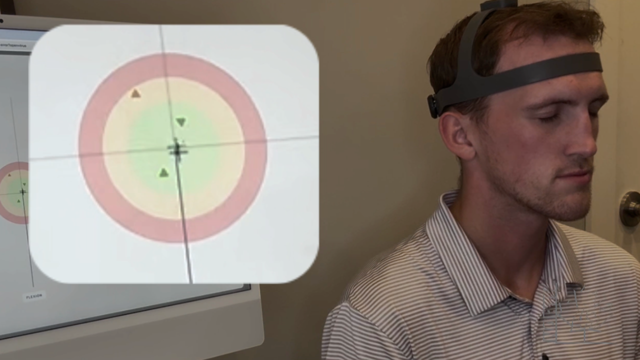
At North 49, we have also appreciated using the NeckCare System as another option for assessing cervical sensorimotor function. It allows us to objectively examine aspects of cervical proprioception and fine motor control that may not be apparent during a traditional musculoskeletal examination.
The Bottom Line
Persistent symptoms after concussion may have multiple contributors, and the cervical spine deserves to be considered among them. Range of motion, pain and palpatory findings matter, but they may not tell us whether cervical proprioception and motor control have recovered.
The question isn't whether the neck should be assessed after concussion. The question is whether we're assessing everything the neck actually does.
References
Author

Kregg Ochitwa
Physical Therapist I Vestibular Therapist I Clinic Owner
Categories
All Categories